Improved test measures virus lethal to young spiny lobsters
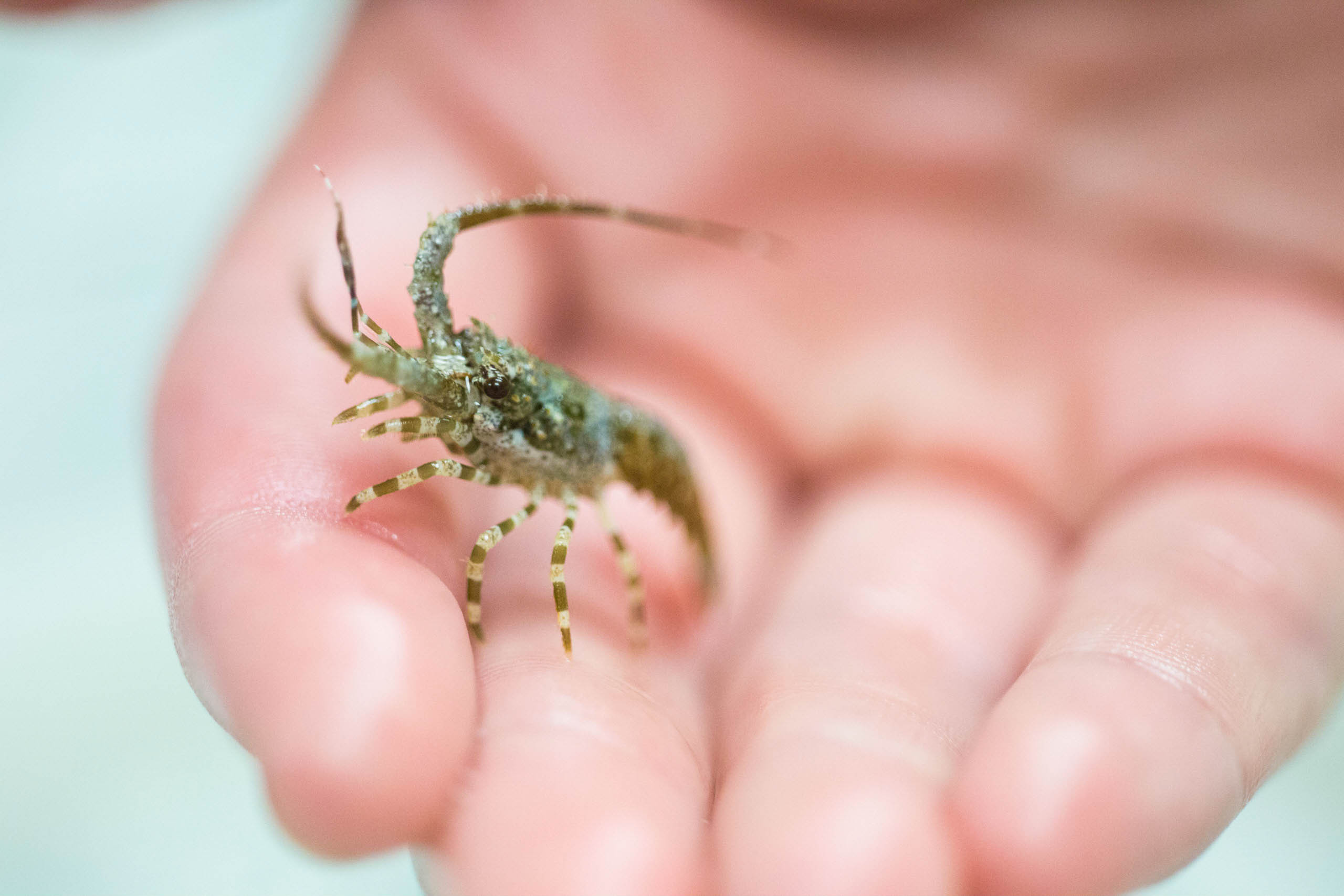
First stage juvenile spiny lobster at Mote Marine Laboratory. Credit: Olivia Raney/Mote Marine Laboratory
A virus that can sicken or kill young Caribbean spiny lobsters can be detected with greater sensitivity — and its abundance can be measured to investigate disease severity and spread — thanks to a new diagnostic test described in a peer-reviewed journal article published recently in Diseases of Aquatic Organisms.
The study to develop this test was led by Mote Marine Laboratory Staff Biologist Dr. Abigail Clark, who began the study while at the University of Florida (UF) with partners from UF and the Virginia Institute of Marine Science (VIMS), and with grants from Florida Sea Grant Scholars, UF Opportunity Seed Fund, and the National Science Foundation.
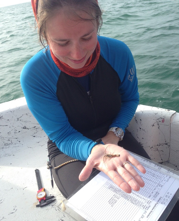
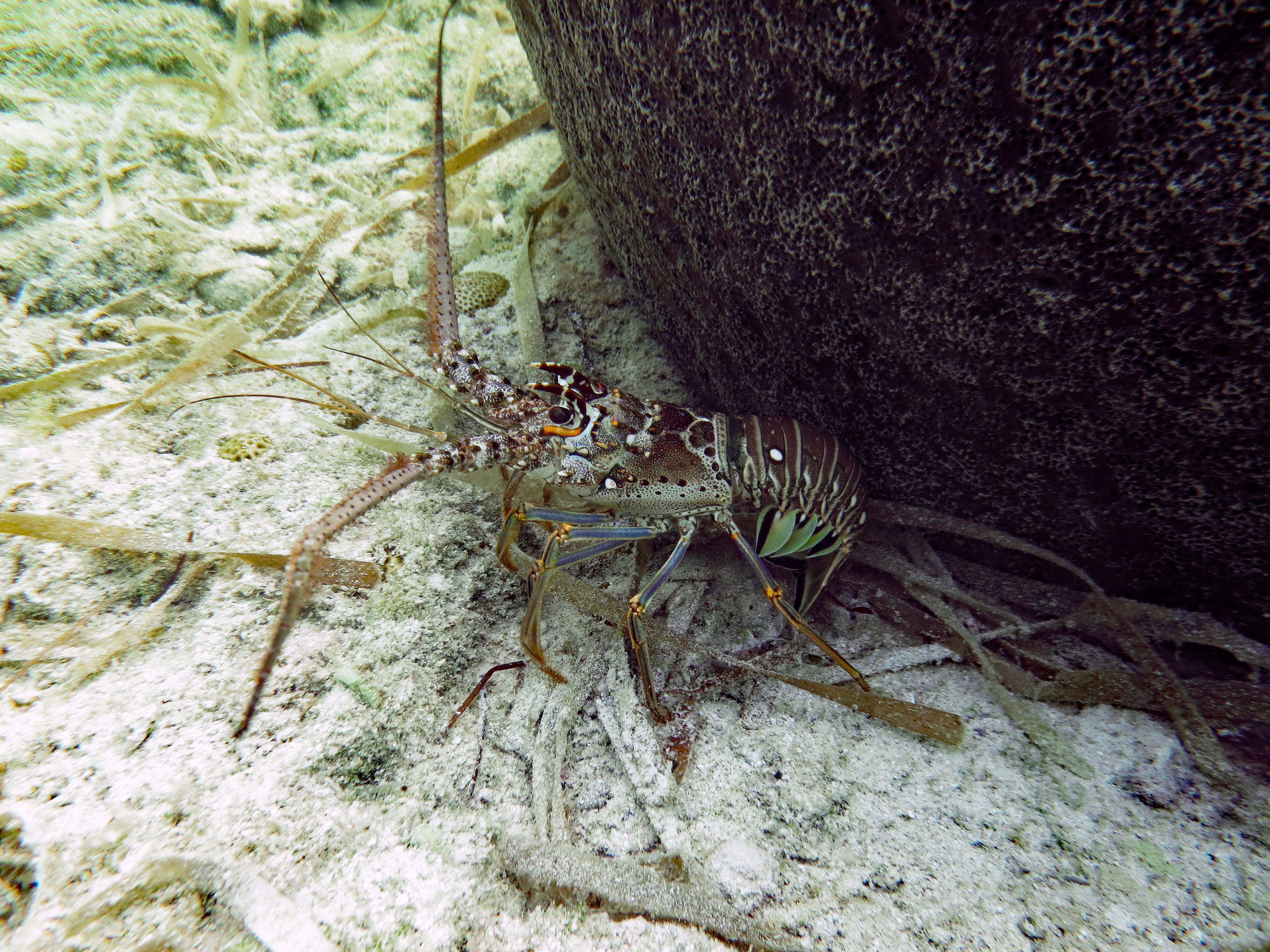
Left: Dr. Abigail Clark holds a juvenile Caribbean spiny lobster. Credit: Alyssa Thompson. Right: Healthy lobster. Credit: Dr. Abigail Clark.
Caribbean spiny lobsters (Panulirus argus) comprise one of the most economically valuable fisheries in their region and are the only known host for the virus, Panulirus argus Virus 1 (PaV1). The virus can kill juvenile lobsters within weeks to months, and symptoms include a discolored shell, hemolymph (blood-like fluid) that looks uncharacteristically “milky,” and lethargic behavior that can leave lobsters vulnerable to predators. However, lobsters of most age groups can carry PaV1 without obvious symptoms of infection. Interestingly, healthy lobsters tend to avoid infected ones — a potential concern for this relatively social species that can be trapped by using a live spiny lobster as bait. Scientists are investigating whether the virus, discovered in 2000, has been affecting fishery yields or might do so with changing, future environmental conditions. So far the answer remains unclear, and studying the virus in detail is an important precaution for managing this valuable fishery.
“We know that PaV1 is present in about 11 percent of spiny lobsters landed in Florida, but adults typically don’t show signs of infection and juveniles might not show signs until they are really, really sick — so we need molecular testing to detect the disease,” said Clark, who continues her research based at Mote’s Elizabeth Moore International Center for Coral Reef Research & Restoration (IC2R3) on Summerland Key, Florida. “A molecular test established by others has allowed us to detect the virus’ DNA in infected hemolymph or tissue, and now we’ve taken this a step further by developing a test that not only detects the virus, but also quantifies the number of viral copies per sample — the viral load — to better understand how sick the lobster is.”
The molecular test confirmed by others is called “endpoint polymerase chain reaction” (endpoint PCR). It involves extracting DNA from lobster tissue samples, using a common method to repeatedly copy one specific stretch of DNA found within the PaV1 virus, making the DNA easier to detect with scientific instruments. Scientists place the copied DNA into electrically charged gel that separates DNA pieces by size and electric charge. Scientists check the gel for what looks like a dash mark, called a band, in the spot where they’d expect the virus DNA to stick. Finding a band in the expected place suggests that the lobster had the virus, though sequencing of the band (reading its “genetic code” using specialized instruments) is required for confirmation.
This test is sensitive and effective at diagnosing lobsters positive or negative for the PaV1 virus. The new study took diagnosis a step further, using another popular and advanced method called “real-time, quantitative PCR” (qPCR), which works somewhat like endpoint PCR but also allows scientists to estimate how many copies of the virus were in the lobster tissue AND detect them even in low numbers.
The current study was the first to test for PaV1 in lobsters using the qPCR method. This method also involves making copies of the viral DNA, but in a new step, scientists add a fluorescent molecule that glows during a certain part of the copying process. By measuring the intensity of that glow, scientists can monitor the number of DNA copies being made in real time, and use that information to estimate how many copies of the virus were in the lobster tissue – indicating how sick the animal is.
Project partners developed this test to check for a specific stretch of DNA in the PaV1 virus, and they examined copies of that DNA from eight regions of the Caribbean Sea, to make sure any regional variation wouldn’t throw off the test. They tried the test with 165 samples of the blood-like hemolymph or leg tissue from juvenile and adult Caribbean spiny lobsters.
The new qPCR test performed just as well as the previous test in detecting the disease – which the scientists learned by taking “positive” samples diagnosed with the past test and running them again with the new one. As hoped, the new qPCR test also allowed scientists to estimate the abundance of the virus in a sample, and repeated testing provided consistent and reliable results. The new test also diagnosed a small number of samples positive after the earlier test had diagnosed them negative. The qPCR method is generally considered more sensitive than endpoint PCR because qPCR is designed to multiply a shorter region of the DNA sequence, which is more efficient than multiplying a longer one.
The researchers also confirmed that their qPCR test would detect the PaV1 virus specifically, and wouldn’t be “fooled” by other viruses found in crustaceans like shrimp and crabs.
For example, Clark said: “We tested a virus found in shrimp, the white spot syndrome virus, against our qPCR assay to make sure it didn’t cross react. The goal was to develop a very specific assay for PaV1. We did find that it was specific — that was awesome.”
With evidence that the qPCR test is highly specific, sensitive, reliable and able to help quantify how severely a lobster is infected, project scientists are excited to apply it in future studies. The test may help scientists better understand how the virus is distributed and progresses within a lobster’s body, and how prevalent it is in seawater and in different populations of Caribbean spiny lobsters — important for understanding how it might spread.
“The better we understand the virus on a molecular level, and understand its presence in the lobsters and their environment, the better opportunities resource managers will have for controlling and managing for it,” Clark said. “One thing worth looking at with qPCR is spatial epidemiology of the virus, and how it may be affected by habitat changes. Many complex factors can influence a disease; for instance, with the tissue-loss disease significantly affecting coral reefs in the Florida Keys now, we don’t fully understand what opened the door for its onset and widespread progression. Even if PaV1 is not damaging the Caribbean spiny lobster fisheries now, we need to keep an eye on it to understand what conditions have the potential to change that.”