Return to Coral Biorepository Alliance Home
Coral Sampling for Genetic Identification
Coral Biorepository Alliance members are working collaboratively to share and standardize methods used to preserve coral genetic diversity for conservation and restoration. Currently, the methods used by our members include sampling, preserving, storing, studying, and restoring coral genetic diversity using three types of biorepositories. Biorepository data for all samples should be held in integrated coral database systems, allowing for parallel and iterative uses of genotypes and samples across biorepository types. Where appropriate, corals should be collected following a free, prior, and informed consent process.
Collecting for Coral Biorepositories – the numbers
The number of individuals/species needed for a biorepository can be calculated using the information in Hagedorn et al. (2025) as described in Box 1.
Box 1: How many organisms per species do we need to collect?
The goals of an ex situ population determine how many genetically distinct organisms per species are needed from the wild as “founders.” One common goal is to insure against complete extinction by maintaining adaptive potential to support reintroductions in the future. Measuring adaptive potential directly is difficult, but it is expected to be correlated with gene diversity (GD), which is straightforward to estimate with molecular tools (Lynch and Walsh 1998). Basic sampling theory indicates that in diploid organisms:
GDf = GDw (1−1/(2Nf))
in which GDf is the gene diversity of the founders, GDw is the gene diversity of the wild population, and Nf is the number of founders (Crow and Kimura 1970). This means that the first ten founders capture a much greater proportion of GDw (95%) than do the second ten (just an additional 2.5%), such that together these first 20 capture 97.5% of GDw (Fig. 2).
For this reason, 20 unrelated founders, sampled widely from within each unit to be conserved, is often used as a benchmark for starting an insurance population. However, this simple rule of thumb presumes that all survive and reproduce, so the actual number of organisms sampled should be somewhat higher to consider likely losses during sampling and variance in future reproductive success (Shearer et al. 2009). Also, it applies only to capturing GD, and not to other measures of genetic diversity, which may require much higher sample sizes (such as retaining rare allelic variants; (Lacy 1994, Baums et al. 2019)).
From a practical standpoint, for example, if there are losses of 50% during sampling, then the initial number of organisms in a species needed would be 40. If subsequent mortality prior to reproduction was 50%, then the initial collection should be 80. If only half those organisms are likely to reproduce, then the number should be 160. Finally, if the only organisms available were closely related, then the number would again need to be increased to compensate. If the goal were to maintain rare allelic variants, rather than to maintain gene diversity, the number would have to be much higher, depending on the rarity of the desired variants.
It may also be useful to evaluate collection completeness for a population by estimating the probability of capturing rare alleles with the following formula (Blackburn, 2009):
Probability of capturing rare alleles = 1 – (1 – A)2N
Where:
A = the allele frequency, and N = the number of animals contributing gametes to the collection.
Therefore, with 40 individuals being sampled, the probability of capturing an allele with a frequency of 0.01 is 55%, and for 80 animals, it is 80%.
Once you have determined how many individuals you want to collect and where, it is important to know if you have the capacity, either in live culture, in your freezer or in a cryo-bank. If you do not have the space, you must adjust your numbers. Moreover, it is always good to have parallel storage for your important samples. Arrange and know your storage limitations before you go out collecting.
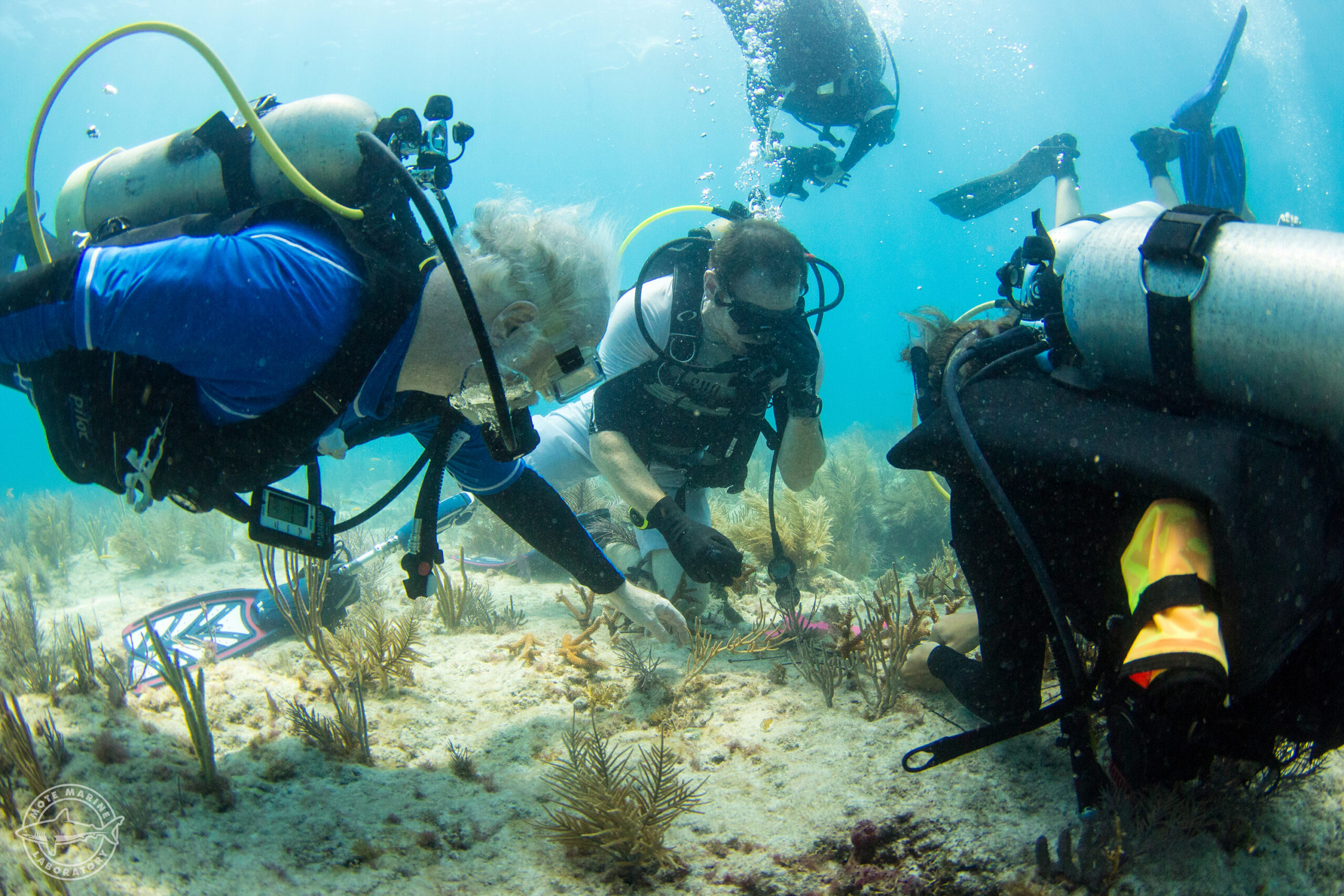
Now, it is time to begin collecting coral fragments or whole colonies. This is best done on scuba.
Collections of fragments:
- Tools needed for coral fragmentation: bone cutter and hole saws and underwater drill (massive corals), hammer, chisel, 70% alcohol
- Understand how large a fragment you will need before you go out based on the types of sub-collections you might want to make for genomics, histology, isotope and voucher samples. If this is the case, you might need ~15 cm piece from each colony.
- Photograph colony in situ from many angles (see details on photographing colonies underwater)
- Take the GPS coordinates (see taking GPS measurements)
- Tag the colony in some way, e.g., cattle tag, so if you have to come back and get additional samples, you can
- Make small tags from underwater paper with unique identifiers that you will attach to your coral. This may be based on a museum system, the date, species, and colony number, etc
- Place the unique identifier tag with the cut fragment in a plastic bag with damp bubble wrap, and place in a cooler
- Clean the bone cutter with alcohol before using it on another colony
- Transport to the lab within a few hours, and place in a water system with good seawater, air and water movement, if needed
- Attach a permanent tag, like an RFID chip, to the colony or fragment (see tagging with RFID chips)
- Enter the data, including the unique identifier number and associated RFID chip, into a database
Step-by-Step Collection Protocol

Before diving or entering the water:
- Prepare and label all collection tubes in advance. Include colony ID, date, site, and intended analysis on each label, linked to a database.
- Pre-fill each tube with the appropriate preservation buffer. For DNA/RNA Shield: 500 to 700 uL per 2 mL tube; 6 mL per 15 mL Falcon tube. For ethanol-only samples: fill to approximately 70% of tube volume (be aware of the volume of your tissue and the need for about 5 to 10 x volume of preservative)
- Place tubes in a cooler with ice or keep at ambient temperature depending on the preservation buffer used.
At the collection site:
- Put on fresh nitrile gloves.
- Identify the target colony and photograph it (in situ, if possible) with a reference marker before sampling.
- For branching corals, position the bone cutters at a branch tip 0.5 to 1 cm from the growing edge and make a single clean cut. For massive corals, place the chisel at the colony margin and strike firmly to detach a fragment of approximately 1 to 2 cm.
- Transfer the fragment immediately into the pre-filled collection tube. Cap securely and

Curacao invert once to ensure contact with the preservation buffer.
- Before moving to the next colony, wipe all cutting surfaces with 70% ethanol and allow to air-dry for at least 30 seconds. If microbiome analyses are planned, follow with a 30-second treatment with 10% bleach, then rinse with sterile water. Change gloves. [4]
After collection:
- Transfer samples to ice, freezer or liquid nitrogen (depending on need) as soon as possible. Samples in DNA/RNA Shield can tolerate ambient temperature for several hours; samples intended for RNA analysis should be chilled within minutes.
- For the -20°C storage target described in this SOP, samples should ideally be frozen within one hour of collection.
- Keep samples frozen until they are processed. If shipping internationally, confirm import permit requirements and carrier dry-ice policies before booking the shipment [original SOP shipping note].
Supplies Needed for Field Collection (Depending on Need)
- Tubes
– Lysing Matrix A tubes, 2 mL (MP Biomedicals, catalog 116930050)
– 2 mL screw-cap O-ring microcentrifuge tubes
– 15 mL polypropylene Falcon tubes
- Preservatives (DNA/RNA Shield (Zymo Research, catalog R1100-250) etc)
- 95% (190-proof) non-denatured ethanol for preservation and tool cleaning
- 10% sodium hypochlorite solution (diluted fresh from household bleach) for tool cleaning
- Sterile water for rinsing tools after bleach treatment
- Bone cutters (rongeurs) for branching corals
- Hammer and chisel for massive and encrusting corals
- Nitrile gloves (multiple boxes; change between each colony)
- Waterproof marker or cryogenic labels for labeling tubes underwater or immediately after surfacing
- A small cooler with ice or, if RNA is a priority, a dry-shipper or liquid nitrogen flask
- A 10 mL pipette and controller for dispensing preservation buffer
- A field balance (scale) capable of measuring to 0.1 g
Key References
[1] Frade PR, Hoey JA, Goodbody-Gringley G, Latijnhouwers KRW, Silberhumer HE, Muir P, Bongaerts P. Hybridization as driving force for cryptic species diversity in the Caribbean coral genus Madracis. Sci Rep. 2025;15:33359. doi:10.1038/s41598-025-17038-6.
[2] Kitchen SA, Von Kuster G, Vasquez Kuntz KL, Reich HG, Miller W, Griffin S, Fogarty ND, Baums IB. STAGdb: a 30K SNP genotyping array and Science Gateway for Acropora corals and their dinoflagellate symbionts. Sci Rep. 2020;10:12488. doi:10.1038/s41598-020-69101-z. PMID: 32719467.
[3] Cowman PF, Quattrini AM, Bridge TCL, Watkins-Colwell GJ, Fadli N, Grinblat M, Roberts TE, McFadden CS, Miller DJ, Baird AH. An enhanced target-enrichment bait set for Hexacorallia provides phylogenomic resolution of the staghorn corals (Acroporidae) and close relatives. Mol Phylogenet Evol. 2020;153:106944. doi:10.1016/j.ympev.2020.106944. PMID: 32860973.
[4] Vega Thurber R, Schmeltzer ER, Grottoli AG, van Woesik R, Toonen RJ, Warner M, et al. Unified methods in collecting, preserving, and archiving coral bleaching and restoration specimens to increase sample utility and interdisciplinary collaboration. PeerJ. 2022;10:e14176. doi:10.7717/peerj.14176. PMID: 36345483.
[5] Wainwright BJ, Zahn GL, Arlyza IS, Amend AS. Comparative methods for preservation and DNA extraction of five taxonomically disparate coral microbiomes. Front Mar Sci. 2021;8:684161. doi:10.3389/fmars.2021.684161.
[6] Voolstra CR, Perna G, Chuard A, Ziegler M. DNA preservation and DNA extraction protocol for field collection of coral samples suitable for host-, marker gene-, and metagenomics-based sequencing approaches. Zenodo. 2022. doi:10.5281/zenodo.6962735.
[7] Voolstra CR, Perna G, Chuard A, Ziegler M. RNA preservation and RNA extraction protocol suitable for field collection of coral samples. Zenodo. 2022. doi:10.5281/zenodo.7108092.
[8] Bergamo TF, Morrow KM, Avila-Magana V, Camp EF, Madin JS, Kutti T, et al. A comparative protocol for preserving deep-water marine invertebrate tissues: DNA/RNA Shield vs. liquid nitrogen for dual extraction of high-quality nucleic acids. Front Mar Sci. 2025. doi:10.3389/fmars.2025.1592143. PMC12645289.
[9] Gray MA, Pratte ZA, Kellogg CA. Comparison of DNA preservation methods for environmental bacterial community samples. FEMS Microbiol Ecol. 2013;83:468-477. doi:10.1111/1574-6941.12008. PMID: 22924961.
[10] van de Water JA, Ainsworth TD, Leggat W, Bourne DG, Willis BL, Tout J. The coral immune response facilitates protection against microbes during tissue regeneration. Mol Ecol. 2015;24:3390-3404. doi:10.1111/mec.13257.
[11] Aranda M, Ziegler M, Voolstra CR. A comparative analysis of microbial DNA preparation methods for use with massive and branching coral growth forms. Front Mar Sci. 2018;5:269. doi:10.3389/fmars.2018.00269. PMC6137167.
[12] Kitchen SA, Von Kuster G, Vasquez Kuntz KL, et al. STAGdb: a 30K SNP genotyping array. Sci Rep. 2020;10:12488 (bioRxiv version, sample quantity guidance). doi:10.1101/2020.01.21.914424.
Why Genetics Matters for Your Coral Collection
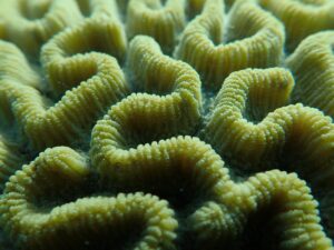
Knowing exactly which species make up a coral collection is essential, especially before using those corals for breeding. In places with relatively few coral species, such as Hawaii, telling species apart by eye is usually straightforward. In other regions, however, corals can interbreed, change shape depending on their environment, or otherwise blur the boundaries between species, which makes visual identification unreliable on its own.
When there is any doubt, genetic testing is the most reliable way to confirm identity. [1] Just as important is labeling every colony and fragment carefully, so that its identity can be tracked over time as it is propagated, fragmented, or moved between facilities. The guidance below is meant as a starting point, not a substitute for working directly with a genetics specialist who can recommend the best method for a specific question.
Matching a Genetic Method to Your Question
The right genetic test depends on the question being asked. The table below summarizes four questions facility managers commonly raise, the method best suited to each one, and an approximate cost per sample.
| Question you are asking | Possible Best Method | Approximate Costs* |
|---|---|---|
| Could the colonies in my tanks be close relatives that might not breed well together?
(A common question in Caribbean restoration work, where keeping genetic diversity high matters.) |
A 30,000-marker SNP genotyping chip built specifically for Acropora corals and their algal symbiotic partners, known as STAGdb. [2] | Provider quotes for a 96-sample plate run from about $76 to $258, depending on the lab and exact assay used. |
| What species do I actually have in my tanks?
(A common question in Australia and other regions with many coexisting coral species.) |
A target-capture sequencing method that reads thousands of shared genetic markers at once, built on a custom Hexacorallia bait set with IDT xGen library preparation chemistry. [3] | About $100 per sample, including DNA extraction, through the Australian Genome Research Facility (AGRF). |
| These colonies look alike, but I am not sure they are really the same species.
(Comes up anywhere sibling species or possible hybrids are suspected.) |
A combined approach pairing reduced-genome sequencing (nextRAD) with detailed shape measurements, microscopy, and life-history comparisons. [1] | Complex and not cheap. Best handled together with a taxonomy and genetics specialist rather than budgeted as a flat per-sample cost. |
| How complex is the microbial community living with my coral? | Bacterial and microbial community genomics, including paired metagenome and metatranscriptome sequencing. | Quoted at roughly 800 EUR (about $852) per sample. |
* These figures do not typically include the cost of DNA extraction or downstream bioinformatics analysis, and they reflect quotes collected directly from providers, which change over time. Anyone budgeting a project should request a current quote from the provider before planning a sampling effort.
How the SNP Genotyping Workflow Works
For groups choosing the SNP array approach, the process generally follows four stages: collecting tissue and extracting DNA, running the sample through the chip and an automated analysis pipeline, receiving a set of deliverables that describe each sample’s genetic identity, and archiving the results in a shared database that other research groups can access. The diagram below illustrates this workflow as implemented in the STAGdb system.

Figure 1. The STAGdb genotyping and data workflow, from sample collection through database archiving. Adapted from Kitchen et al., 2020, Scientific Reports, used under a Creative Commons Attribution 4.0 license.
Field Sampling Protocol for Coral Genetics
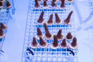
This protocol covers the methods, equipment, and storage conditions needed to collect coral tissue in the field for DNA and RNA applications. The amount of tissue required depends on the downstream genetic process; these requirements, and the evidence behind them, are described in the sections that follow.
Sampling Methods
Three physical approaches are used to obtain coral tissue in the field, each suited to different situations. [4]
Bone cutters (rongeurs). For branching corals such as Acropora and Pocillopora, bone cutters are the most common and least disruptive tool. A single cut through a branch tip of 0.5 to 2 cm yields sufficient material for most genetic applications. [6,7] Bone cutters work poorly on massive or encrusting species and should not be forced, as this risks shattering the tissue rather than making a clean cut.
Hammer and chisel. For massive and encrusting corals such as Porites and Orbicella, a hammer and chisel are more reliable. A fragment of roughly 1 to 2 cm in the shortest dimension is generally sufficient. The chisel is placed at the colony margin where tissue is thicker and more easily removed from the skeleton, then struck firmly to produce a clean break. [4]
Airbrushing tissue from the skeleton. In laboratory or vessel settings where a pressurized air supply is available, coral tissue can be removed from a frozen or fresh fragment by directing a fine stream of air or phosphate-buffered saline at the surface. The resulting tissue slurry is collected and centrifuged to separate cells from the overlying fluid. This method yields a cleaner separation of host tissue from the skeleton and is well suited to downstream microbial and metagenomic work because it reduces contamination from endolithic organisms living inside the skeleton. [11]
How Much Tissue Is Needed for Each Genetic Process
The table below summarizes the tissue input requirements for each genetic method used in this project. These figures reflect the literature rather than arbitrary defaults; the key references are noted in the right column.
| Genetic Process | Recommended Tissue Input | Notes | Key References |
|---|---|---|---|
| SNP genotyping array (STAGdb) | 3 to 4 polyps (approximately 50 to 100 mg wet tissue), yielding DNA at 63 pg/µL or higher | Higher concentrations improve call rate. Even moderately degraded DNA is acceptable for this platform. 95% ethanol is the preferred preservative. | Kitchen et al. (2020); Vega Thurber et al. (2022); Voolstra et al. (2023) |
| Target-capture sequencing (Hexacorallia bait set) | 600 ng total genomic DNA (at 10 ng/µL), derived from approximately 0.5 to 1 cm of branch or 100 to 200 mg of tissue | DNA is sheared to 400 to 800 bp before library preparation. Clean, high-molecular-weight DNA with a 260/280 ratio of 1.8 to 2.1 is required. | Cowman et al. (2020); Vega Thurber et al. (2022) |
| nextRAD / reduced-representation sequencing | At least 300 ng total DNA, derived from approximately 50 to 200 mg of tissue | Quality requirements are similar to the SNP array. Animal tissue input of 0.5 g or more is recommended to ensure adequate yield. | Russello et al. (2015); Vega Thurber et al. (2022) |
| Microbial community / metagenomics (16S, ITS2, shotgun) | 0.5 to 1 cm branch tip or 0.17 to 0.25 g whole coral fragment (tissue plus skeleton), transferred to a bead-beating tube | Bead-beating homogenization is required. Over-loading the tube reduces yield, particularly in mucus-rich species. Separate extraction kits are used for host DNA versus microbial DNA. | Vega Thurber et al. (2022); Silva et al. (2022); Belser et al. (2023) |
| RNA extraction / transcriptomics | Approximately 300 mg of fresh tissue in 1 mL preservation buffer; or a 0.5 to 1 cm branch tip placed immediately into buffer | RNA is far more labile than DNA. The tissue must contact preservation buffer within seconds of collection. Cold-water coral species require liquid nitrogen; tropical species can be preserved with DNA/RNA Shield if processed promptly. | Vega Thurber et al. (2022); Mayfield et al. (2016); Belser et al. (2023) |
| Metatranscriptomics (holobiont) | 0.5 to 1 cm branch tip or equivalent, processed using a combined DNA/RNA extraction protocol
(Same fragment as for metagenomics if coordinated.) |
RNA integrity number (RIN) of at least 8 is the target for this application. A bead-beating step before extraction is required. | Voolstra et al. (2015); Bernal-Morales et al. (2023); Vega Thurber et al. (2022); Belser et al. (2023) |
References for Tissue Requirements Table
Kitchen et al. (2020): Kitchen SA, Von Kuster G, Vasquez Kuntz KL, Reich HG, Miller W, Griffin S, Fogarty ND, Baums IB. STAGdb: a 30K SNP genotyping array and Science Gateway for Acropora corals and their dinoflagellate symbionts. Sci Rep. 2020;10:12488. doi: 10.1038/s41598-020-69101-z. PMID: 32719467.
Cowman et al. (2020): Cowman PF, Quattrini AM, Bridge TCL, Watkins-Colwell GJ, Fadli N, Grinblat M, Roberts TE, McFadden CS, Miller DJ, Baird AH. An enhanced target-enrichment bait set for Hexacorallia provides phylogenomic resolution of the staghorn corals (Acroporidae) and close relatives. Mol Phylogenet Evol. 2020;153:106944. doi: 10.1016/j.ympev.2020.106944. PMID: 32860973.
Russello et al. (2015): Russello MA, Waterhouse MD, Etter PD, Johnson EA. From promise to practice: pairing non-invasive sampling with genomics in conservation. PeerJ. 2015;3:e1106. doi: 10.7717/peerj.1106. PMID: 26244119. [Note: Establishes the nextRAD library preparation protocol used by SNPsaurus and cited widely in coral reduced-representation sequencing studies.]
Vega Thurber et al. (2022): Vega Thurber R, Schmeltzer ER, Grottoli AG, van Woesik R, Toonen RJ, Warner M, et al. Unified methods in collecting, preserving, and archiving coral bleaching and restoration specimens to increase sample utility and interdisciplinary collaboration. PeerJ. 2022;10:e14176. doi: 10.7717/peerj.14176. PMID: 36345483.
Silva et al. (2022): Silva DP, Epstein HE, Vega Thurber R. Best practices for generating and analyzing 16S rRNA amplicon data to track coral microbiome dynamics. Front Microbiol. 2022;13:1007877. doi: 10.3389/fmicb.2022.1007877. PMID: 36590416.
Mayfield et al. (2016): Mayfield AB, Wang YB, Chen CS, Chen SH, Lin CY. Compartment-specific transcriptomics in a reef-building coral exposed to elevated temperatures. Mol Ecol. 2016;25(11):2599–2615. doi: 10.1111/mec.13628. PMID: 27027254.
Voolstra et al. (2015): Voolstra CR, Sunagawa S, Matz MV, Bayer T, Aranda M, Buschiazzo E, DeSalvo MK, Lindquist E, Szmant AM, Coffroth MA, Medina M. Rapid evolution of coral proteins responsible for interaction with the environment. PLoS One. 2011;6(5):e20392. doi: 10.1371/journal.pone.0020392. PMID: 21647456. [Note: Voolstra and colleagues established metatranscriptomic holobiont protocols including combined DNA/RNA extraction and the RIN ≥8 quality standard; see also Voolstra CR, et al. Front Mar Sci. 2015;2:62. doi: 10.3389/fmars.2015.00062.]
Bernal-Morales et al. (2023): Bernal-Morales E, Enríquez S, Aguayo-Pecina A, González-Pech RA, Iglesias-Prieto R. Integrative omics framework for characterization of coral reef ecosystems from the Tara Pacific expedition. Sci Data. 2023;10:334. doi: 10.1038/s41597-023-02204-0. PMID: 37264047. [Note: This study established RNA integrity number (RIN) of at least 8 as the quality target for holobiont metatranscriptomics and confirmed the bead-beating combined extraction protocol for coral tissue.]
Voolstra, C.R.et al. (2023). Disparate genetic divergence patterns in three corals across a pan-Pacific environmental gradient highlight species-specific adaptation. npj Biodiversity 2, 15. https://doi.org/10.1038/s44185-023-00020-8.
Belser, C et al. (2023). Integrative omics framework for characterization of coral reef ecosystems from the Tara Pacific expedition. Sci Data 10, 326. https://doi.org/10.1038/s41597-023-02204-0.
Collection Tubes and Containers
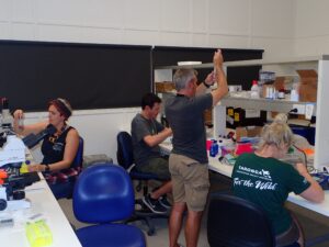
The choice of collection tube affects both DNA and RNA quality and must match the downstream application.
Lysing Matrix A tubes (MP Biomedicals, catalog 116930050). These 2 mL tubes contain ceramic beads and are the primary collection vessel when bead-beating homogenization will follow. They are used for a fragment of 0.5 to 1 cm placed in 700 uL of the appropriate lysis or preservation buffer. They are compatible with a range of extraction kits including the Qiagen AllPrep DNA/RNA Mini kit. [11]
2 mL screw-cap microcentrifuge tubes with O-ring seals. For samples stored in 95% ethanol or DNA/RNA Shield without immediate homogenization, a standard 2 mL O-ring tube is adequate and reduces the risk of leakage during shipping. O-ring tubes are preferred over standard snap-cap tubes for shipments involving temperature changes. [4,5]
15 mL polypropylene Falcon tubes. A 15 mL Falcon tube is used when a larger fragment (up to 4 g) is collected for potential downstream applications requiring more starting material, such as multiple parallel extractions. The fragment is submerged in 6 mL of DNA/RNA Shield. Polypropylene construction avoids leaching of plasticizers that can inhibit downstream enzymatic steps.
Cryovials. For samples intended for long-term archiving at -80 degrees C, 1 to 2 mL cryovials with external threading are appropriate. They are rated for liquid nitrogen temperatures and resist cracking during freeze-thaw cycles. [4]
Sterilizing Bone Cutters and Other Tools Between Samples
Cross-contamination between colonies is one of the most common and underappreciated sources of error in coral genetic studies and particularly affects microbiome analyses. Aseptic technique requires only minor changes to the standard collection workflow but substantially expands the range of analyses that can be performed on archived tissue. [4]
Between-sample decontamination protocol for bone cutters, chisels, and forceps:
- Rinse cutting surfaces with freshwater or seawater to remove gross tissue debris.
- Submerge or wipe the cutting surface with 70% ethanol and allow to air-dry for a minimum of 30 seconds. Ethanol at 70% is effective against bacteria and most viruses and is standard practice in the coral field. [4]
- Where microbiome integrity is a priority, follow the ethanol step with a brief (30-second) wipe using 10% sodium hypochlorite (household bleach), then rinse with sterile water and allow to dry. This two-step sequence removes both organic contamination and residual microbial DNA from the tool surface. [4]
- Change to a fresh pair of nitrile gloves between each colony. Gloves are a more commonly overlooked source of cross-contamination than tools.
At minimum, 70% ethanol between each sample is required. If microbiome analyses are planned for any samples in the set, apply the full bleach-plus-ethanol sequence throughout the entire collection effort, not only for individual samples of interest.
Preservation Buffers, Storage Temperatures, and Sample Stability
The preservation approach used at the moment of collection determines which downstream analyses are possible. The table below summarizes the most commonly used options, their storage temperature requirements, and the time windows over which sample quality is maintained.
| Preservative | Storage Temperature | Stability Window | Suitable Analyses | Notes |
|---|---|---|---|---|
| DNA/RNA Shield (Zymo Research) | Ambient (field), then −20°C | Hours to days at ambient; months at −20°C | DNA, RNA (tropical corals), 16S, ITS2, metagenomics | Lytic buffer inactivates nucleases and pathogens on contact. Not recommended for RNA in cold-water coral species, where RNA integrity was lower than with liquid nitrogen. [8] |
| 95% Ethanol | Room temperature to −20°C | Years at −20°C; decades possibly based on accelerated aging data | DNA only; genotyping, SNP arrays, 16S, ITS2 | The most commonly used field preservative. Ethanol-stored samples at 40°C for 20 months yielded DNA suitable for host and symbiont genotyping. [9] RNA is not recoverable. |
| RNAlater (Thermo Fisher Scientific) | Room temperature (up to 1 week), 4°C (up to 1 month), −20°C (indefinitely) | Stable indefinitely at −20°C; partial RNA degradation reported at 37°C after 3 days | DNA and RNA; transcriptomics, genotyping | Tissue must be trimmed to less than 0.5 cm in one dimension and submerged immediately in 5× the tissue volume of RNAlater. Compatible with most downstream RNA isolation kits. [7] |
| Liquid Nitrogen / −80°C | Liquid nitrogen in the field; −80°C in the laboratory | Years at −80°C; 7–10 years documented for DNA and RNA integrity | All omics: genomics, transcriptomics, proteomics, metagenomics, metatranscriptomics | The gold standard for RNA and any omics application. Flash freezing is optimal but requires access to liquid nitrogen in the field, which is often impractical at remote reef sites. [4,8] |
| DMSO-Saturated Salt Solution (SSD) | Room temperature to −20°C | Days to weeks at room temperature; longer at −20°C | DNA; PCR, genotyping, 16S | Shown to yield higher molecular-weight DNA and better PCR success than ethanol in some coral species. EDTA in some formulations can leach calcium from the skeleton with prolonged storage. [9] |
Approximate Cost Considerations (Change Constantly)
Fieldwork and laboratory costs interact in ways that are easy to underestimate. The preservation method chosen in the field has direct consequences for downstream costs.
Shipping. Samples frozen at -20 or -80°C require dry ice for shipment. A standard overnight dry-ice shipment within the continental United States typically costs $100 to $300 depending on carrier and volume, and international shipments involving biological materials require permits and additional carrier surcharges. Samples preserved in 95% ethanol or DNA/RNA Shield can be shipped at ambient temperature as non-hazardous biological material if not frozen, substantially reducing freight costs. However, the carrier must be consulted regarding any flammability classification of ethanol-preserved samples. [4]
Extraction. Commercial extraction kits cost roughly $2 to $6 per sample for standard DNA kits (Qiagen DNeasy) and $8 to $15 per sample for combined DNA/RNA kits (Qiagen AllPrep, Zymo Quick-DNA/RNA). Metagenomics-grade kits with differential lysis steps cost more, typically $15 to $30 per sample, not including labor.
Sequencing and genotyping. Sequencing costs are described in the methods table above and are subject to change; always request current quotes before planning a sampling campaign. For the STAGdb SNP array, the cost per sample decreases substantially when a full 96-well plate is submitted, making it worthwhile to coordinate sample collection to fill plates rather than sending partial plates. [2] For target-capture sequencing through AGRF, the roughly $100 per-sample estimate cited in the methods table includes extraction but not bioinformatics. [3] Microbial community metatranscriptomics is the most expensive application at approximately 800 EUR per sample and requires extensive bioinformatics expertise [see original SOP methods table].
Genetic Analyses
These analyses will be done at a number of places:
- Genoscope (Europe) (https://www.cea.fr/drf/ig/english/Pages/Genoscope.aspx)
- AGRF (Australia)(https://www.agrf.org.au/)
- Hawaii Institute of Marine Biology – University of Hawaiʻi (https://www.himb.hawaii.edu/)
- Mote Marine Laboratory – Florida coral restoration genomics (https://mote.org/research/centers-of-excellence/international-coral-gene-bank/)
- Smithsonian National Museum of Natural History – coral phylogenomics (https://naturalhistory.si.edu/)
- Scripps Institution of Oceanography – coral microbiome and genomics (https://scripps.ucsd.edu)